Diagnostic Services
X-Rays & 3D Imaging
Diagnostic imaging lets us see what a clinical exam cannot, helping us catch issues early and plan treatment with confidence.
Why dental imaging matters
A clinical exam is essential, but it only shows the dentist what is visible to the eye. Dental x-rays and 3D imaging reveal what's happening between teeth, below the gumline, and inside the surrounding bone, areas where problems can develop silently before any symptoms appear.
Early detection of decay, bone changes, cysts, or other abnormalities means simpler, less involved treatment. Imaging is one of the most important diagnostic tools available to your dental team.
Digital x-rays
Able Family Dental uses digital x-ray systems, which use significantly less radiation than older film-based technology, typically 70-90% less. The images appear instantly on screen, where they can be enlarged, adjusted, and reviewed with you in the chair.
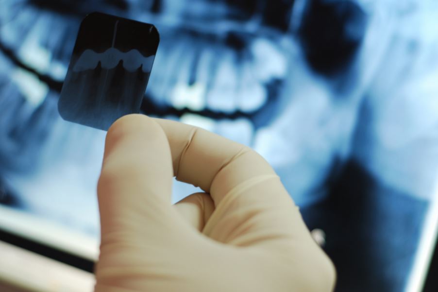
Bitewing x-rays show the crowns of the upper and lower back teeth and are used primarily to detect cavities between teeth and assess bone levels.
Periapical x-rays capture a full tooth from crown to root tip, including the surrounding bone. They are useful for diagnosing abscesses, root fractures, and bone changes around specific teeth.
How often x-rays are taken depends on your individual history, risk level, and how long it has been since the last set. We follow evidence-based guidelines and do not take x-rays on a fixed schedule for everyone, the frequency is personalised.
Panoramic x-rays
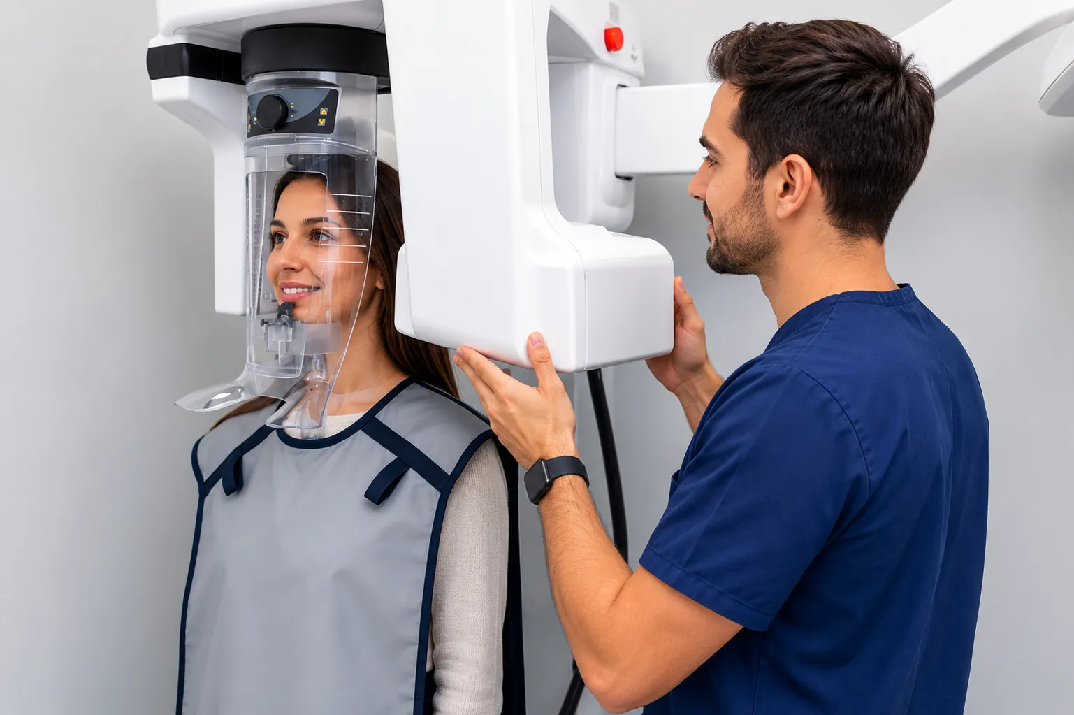
A panoramic x-ray (also called a panorex) captures the entire mouth in a single image, all teeth, both jaws, the temporomandibular joints, and surrounding anatomy. It is commonly taken for new patients to establish baseline records, and periodically thereafter to monitor changes over time.
Panoramic images are also useful for assessing the position of wisdom teeth, evaluating bone levels across the full arch, and identifying abnormalities that may not be visible on smaller films.
Cone beam CT (CBCT) for complex planning
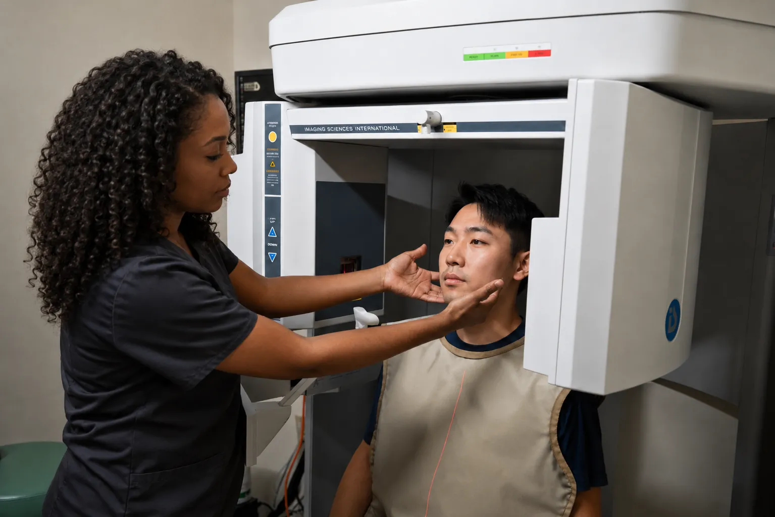
Cone beam computed tomography (CBCT) uses a cone-shaped beam to capture a three-dimensional image of the teeth, jaws, bone, and surrounding anatomy. Unlike a conventional x-ray, a CBCT scan can be viewed in cross-section from any angle, allowing detailed assessment of bone volume and density, nerve pathways, and the precise positioning of teeth.
CBCT is not part of routine dental care. It is ordered when three-dimensional information is necessary for treatment planning. Common situations include dental implant planning, assessment of impacted teeth, complex root canal anatomy, and evaluation of jaw or bone pathology.
A CBCT scan delivers more radiation than a standard dental x-ray, so it is used only when the clinical benefit justifies it. The scan itself is non-invasive, typically takes under a minute, and involves no discomfort.
Ready to get started?
Call 403.327.7202 or request an appointment online.